Diagnostic Ultrasound
Introduction to diagnostic ultrasound
Diagnostic Ultrasound
Diagnostic ultrasound, also known as sonography, is a noninvasive imaging technique that utilizes high-frequency sound waves to produce real-time images of internal body structures. Unlike X-rays or CT scans, ultrasound does not use ionizing radiation, making it a safer option, especially for sensitive groups like pregnant women and children.
Ultrasound is widely used across various medical specialties, including:
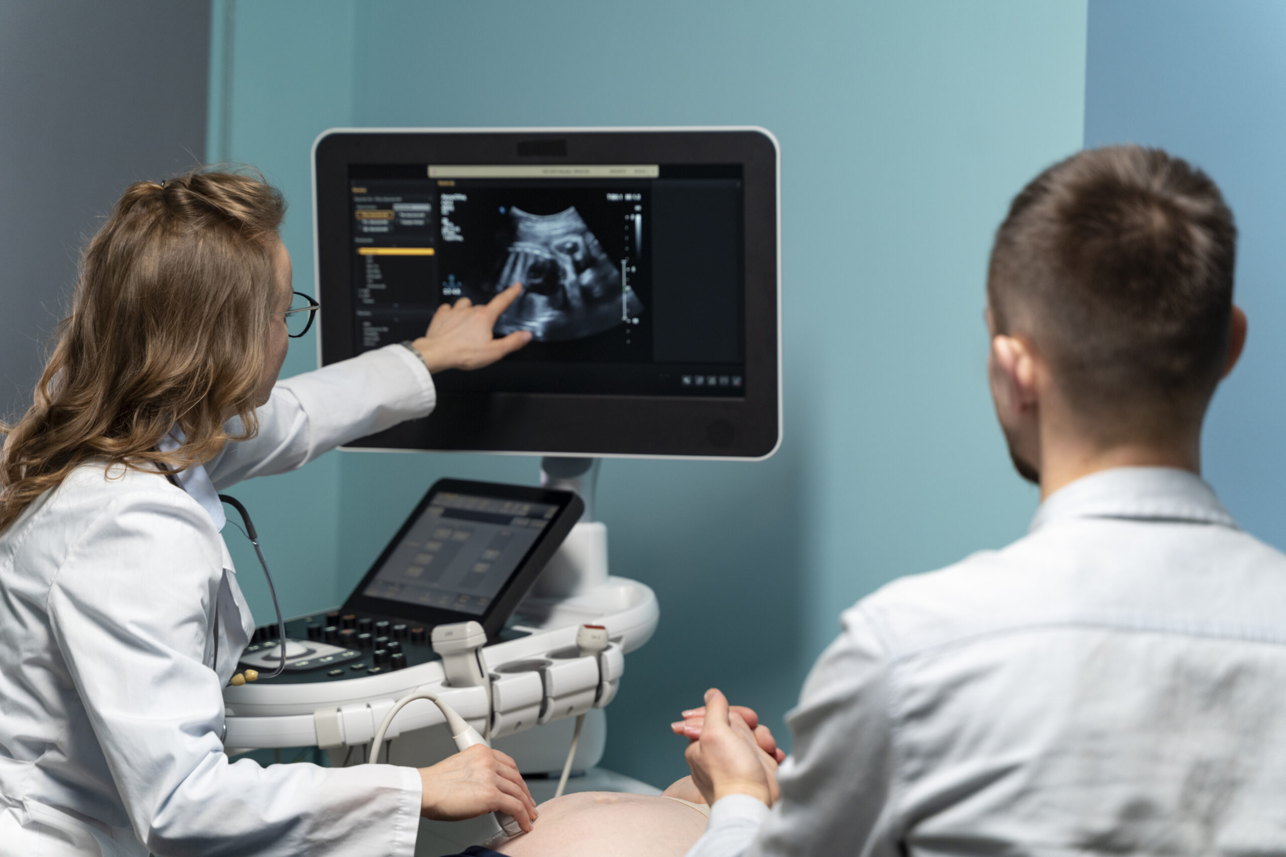
Obstetrics: Monitoring fetal development, detecting abnormalities, and evaluating placental health.
Cardiology: Assessing heart function, measuring blood flow, and visualizing the heart’s chambers and valves.
Musculoskeletal: Imaging muscles, tendons, and joints to diagnose injuries like sprains, tears, and inflammation.
Abdominal Imaging: Ultrasound is commonly used to visualize the liver, kidneys, and gallbladder. It’s a non-invasive tool for diagnosing liver disease, kidney stones, and gallstones. (source)
Vascular: Used for evaluating blood flow, detecting blockages, and diagnosing conditions like DVT and PAD. Doppler ultrasound helps assess blood circulation in veins and arteries. (source)
Thyroid: Ultrasound assists in evaluating thyroid nodules and distinguishing between benign and malignant masses. It’s key in guiding biopsies for further examination. (source)
Breast: Ultrasound helps differentiate between benign and malignant breast lesions, providing valuable support in interpreting mammography results. It’s also used to guide biopsies. (source)
Procedure Guidance: Ultrasound provides real-time imaging for guiding procedures such as biopsies, injections, and fluid drainage, ensuring precision and reducing risks. (source)
What to Look for in a Competitive Product
Image Resolution: Higher-resolution systems will provide more detailed and precise images.
Portability: Portability is essential if you need ultrasound in different settings, such as emergency rooms or remote areas.
Ease of Use: Look for intuitive, user-friendly systems with simple interfaces and adjustable settings.
Advanced Features: Doppler imaging for analyzing blood flow or 3D imaging for obstetrics can add significant value.
Practical Tip
Ensure the system can accommodate multiple diagnostic needs, making it a versatile tool across various specialties.
Science of Diagnostic Ultrasound
Ultrasound Mechanism
Ultrasound emits high-frequency sound waves, typically 2 to 18 MHz, through a transducer. These sound waves interact with tissues, and the resulting echoes are captured by the transducer and transformed into visual images.
Different tissues reflect sound waves to varying degrees, which allows the ultrasound machine to distinguish between muscle, fat, and organs.
Real-Time Imaging and Interpretation of Ultrasound Data
Ultrasound’s most significant advantage is its ability to provide real-time images. This feature is particularly valuable for guiding procedures like needle biopsies, injections, and surgeries. With real-time imaging, clinicians can make immediate decisions during procedures, improving precision and outcomes.
What to Look for in a Competitive Product:
Choose systems that deliver high-quality resolution for still images and video. Additionally, look for color Doppler imaging and tissue elasticity mapping for precise information, especially in specialties like cardiology and oncology.
Real-World Example
In cardiology, high-quality Doppler ultrasound systems can precisely assess blood flow patterns, helping clinicians diagnose heart diseases, valve issues, and congenital disabilities in infants.
Historical Development of Diagnostic Ultrasound
Evolution from Early Diagnostic Tools to Modern High-Resolution Imaging Systems
Ultrasound technology began in the 1950s with large, basic machines that detect tissue abnormalities. Over time, advancements introduced higher-frequency sound waves, Doppler imaging, and 3D/4D capabilities, significantly improving resolution and diagnostic accuracy.
Key Advancements in Portability and Precision
Developing portable ultrasound systems was a game-changer, particularly in emergency and point-of-care settings.
Handheld ultrasound units have enabled imaging in nontraditional settings, such as ambulances and remote areas. They provide quick access to diagnostic imaging without requiring patients to be transported to a hospital for treatment.
What to Look for in a Competitive Product
When selecting ultrasound equipment, consider portability and image precision. Modern ultrasound systems are available in various sizes, ranging from full-sized hospital units to portable, handheld devices. Select the one that balances high-quality imaging and ease of transport, primarily if you work in mobile or urgent care settings.
Battery Life:
Ensure the ultrasound device has long-lasting battery life, especially for portable or handheld units in mobile or urgent care settings. A longer battery life enables continuous usage without frequent recharging, enhancing efficiency.
Artificial Intelligence (AI):
Look for ultrasound equipment that integrates AI for enhanced imaging capabilities. AI can assist in automatic image analysis, improve diagnostic accuracy, and streamline workflow by reducing the need for manual interpretation.
DICOM (Digital Imaging and Communications in Medicine)
Ensure the ultrasound system supports DICOM compatibility for easy integration into hospital or clinic systems. DICOM enables the seamless storage, retrieval, and sharing of medical images, ensuring efficient patient data management.
Latest Research
A recent study in the Journal of Ultrasound in Medicine highlighted the importance of portable ultrasound machines in enhancing access to healthcare in low-resource regions while maintaining diagnostic accuracy.
Mechanisms of Action
How Ultrasound Waves Penetrate Tissues and Create Visual Images
When ultrasound waves are transmitted into the body, they travel through tissues and reflect, depending on the tissue type. The ultrasound system processes these returning echoes to generate detailed images visually representing internal structures. Differences in tissue density, such as between solid organs, muscles, and fluids, help form clear images.
Importance of Doppler Ultrasound for Assessing Blood Flow
A Doppler ultrasound is a specialized technique to measure blood flow within vessels. By tracking the frequency shift of sound waves bouncing off moving red blood cells, Doppler ultrasound provides critical information about the speed and direction of blood flow, essential for diagnosing conditions such as arterial blockages and venous insufficiencies.
What to Look for in a Competitive Product:
Choose a system with advanced Doppler features. High-end ultrasound systems offer color Doppler imaging, which visually color-codes blood flow, making it easier to interpret results quickly.
Real-World Example: Doppler Ultrasound in Obstetrics
Doppler ultrasound is commonly used in obstetrics to monitor blood flow in the umbilical cord, assess fetal health, and detect complications related to placental circulation. Here are patient case studies that demonstrate how Doppler ultrasound is applied in clinical settings to ensure the health of both mother and baby.
Case Studies
Case Study 1: Obstetric Case Study – Monitoring Blood Flow in the Umbilical Cord
Patient Profile:
- Name: Rachel
- Age: 32
- Gender: Female
- Medical History: First-time pregnancy, no significant medical history.
- Gestational Age: 28 weeks
Treatment Process
Rachel underwent a routine ultrasound to monitor fetal health. The obstetrician recommended a Doppler ultrasound due to her family history of hypertension. During the procedure, high-frequency sound waves were used to measure the blood flow through the umbilical cord. The results were analyzed to assess the speed and direction of blood flow, ensuring that the fetus received adequate oxygen and nutrients.
Outcomes
Week 1: The Doppler ultrasound revealed normal blood flow in the umbilical cord, indicating that the fetus was receiving sufficient blood supply.
Follow-Up: At the 32-week follow-up, a subsequent Doppler ultrasound revealed no signs of placental insufficiency or abnormal blood flow, confirming that both mother and baby were doing well.
Findings
The Doppler ultrasound effectively monitored Rachel’s pregnancy, ensuring the proper functioning of the umbilical cord and placental circulation. The procedure provided peace of mind and confirmed no immediate concerns for fetal health.
Case Study 2: Obstetric Case Study – Assessing Fetal Health and Placental Circulation
Patient Profile:
- Name: Laura
- Age: 36
- Gender: Female
- Medical History: Previous miscarriage, high-risk pregnancy due to age, and underlying conditions, including gestational diabetes.
- Gestational Age: 24 weeks
Treatment Process
Laura’s pregnancy was classified as high-risk due to her medical history and age. To assess the health of her fetus and the function of the placenta, a Doppler ultrasound was performed. The procedure measured the blood flow through the umbilical cord and the uterine arteries, evaluating the blood supply to the placenta and fetal organs.
Outcomes
Week 1: The Doppler ultrasound showed normal blood flow to the fetus and placenta, with no signs of restricted blood flow or fetal distress.
Follow-Up: At 30 weeks, another Doppler ultrasound revealed that the placental blood flow remained optimal, a positive indicator of continued fetal health.
Final Outcome: By 38 weeks, Laura successfully delivered a healthy baby, and the Doppler ultrasound provided vital information throughout the pregnancy, ensuring no complications arose from placental or fetal circulation issues.
Findings
The Doppler ultrasound played a crucial role in monitoring Laura’s high-risk pregnancy. It helped track placental health and fetal well-being, enabling early intervention if necessary and ensuring a successful pregnancy outcome.
Case Study 3: Obstetric Case Study – Detecting Placental Complications
Patient Profile:
- Name: Megan
- Age: 40
- Gender: Female
- Medical History: History of preeclampsia in a previous pregnancy, currently pregnant with twins.
- Gestational Age: 26 weeks
Treatment Process
Given Megan’s history of preeclampsia, her obstetrician ordered a Doppler ultrasound to assess placental circulation and monitor fetal health. The procedure focused on measuring blood flow through the umbilical cords of both babies and the uterine arteries to detect early signs of complications, such as intrauterine growth restriction (IUGR) or preeclampsia-related placental insufficiency.
Outcomes
Week 1: The Doppler ultrasound results showed reduced blood flow in the uterine artery, indicating a slight risk of developing preeclampsia. However, the blood flow to the fetuses was within normal ranges.
Follow-Up: At 30 weeks, a second Doppler ultrasound was performed. The results showed that the blood flow to Baby A was slightly restricted, prompting further monitoring and adjustments to Megan’s care plan.
Final Outcome: By 36 weeks, the second baby was born without complications, but Baby A required a brief stay in the neonatal intensive care unit (NICU) for monitoring. The Doppler ultrasound enabled the early identification of potential complications, facilitating timely interventions.
Case Study 4: Ultrasound-Guided Acromioclavicular (AC) Joint Injection for Shoulder Pain Relief
Patient Profile:
- Age: 65
- Occupation: Manual laborer
- Gender: Female
- Medical History: Persistent right shoulder pain unresponsive to conservative treatments
Clinical Presentation
The patient presented with chronic right shoulder pain, mainly localized over the AC joint. Despite undergoing physical therapy and taking nonsteroidal anti-inflammatory drugs (NSAIDs), she experienced minimal relief. The pain significantly impacted her ability to perform daily tasks, particularly those that required overhead movements.
Diagnostic Approach
Physical examination revealed tenderness over the AC joint and limited shoulder range of motion. Imaging studies, including X-rays, confirmed signs of AC joint arthritis.
An ultrasound examination was performed to further assess the condition and guide treatment. The examination identified inflammation and degeneration within the AC joint.
Treatment Intervention
An ultrasound-guided injection of the AC joint was administered, consisting of a local anesthetic and corticosteroid to reduce inflammation and alleviate pain. Using ultrasound guidance ensured precise needle placement, enhancing the effectiveness of the injection and minimizing potential complications. NYSORA
Outcomes
One month after the injection, the patient reported pain relief and had regained full shoulder mobility. She returned to her manual labor job without discomfort and experienced no adverse effects from the procedure.
Conclusion
This case demonstrates the efficacy of ultrasound-guided AC joint injections in providing significant pain relief and functional improvement for patients with AC joint arthritis who are unresponsive to conservative treatments. The precision offered by ultrasound guidance significantly contributes to the success and safety of the procedure.
Therapeutic Benefits
Diagnostic Capabilities in Detecting Tumors, Soft Tissue Injuries, and Organ Abnormalities
Diagnostic ultrasound is invaluable for detecting tumors, soft tissue injuries, and organ abnormalities.
It is commonly used to examine the liver, kidneys, and heart and to detect tumors, cysts, or abnormalities in muscles, tendons, and joints. Ultrasound is crucial for early tumor detection and guiding biopsies, ensuring that tissue samples are collected accurately and precisely.
Guiding Therapeutic Procedures Like Injections and Biopsies
Ultrasound-guided procedures, including injections, fluid drainage, and biopsies, benefit greatly from real-time imaging. This increases the precision of these procedures, reducing the risk of complications and ensuring accurate needle or catheter placement.
What to Look for in a Competitive Product:
When selecting an ultrasound system, ensure it provides high-resolution images that guide therapeutic interventions. Features like needle guidance, which overlays the needle path on real-time images, can increase procedural accuracy.
Benefits and Outcomes:
For patients undergoing biopsies, real-time imaging ensures that the tissue sample is taken from the correct location, enhancing diagnostic accuracy and reducing the need for repeat procedures.
Competitor Comparison
How Diagnostic Ultrasound Compares to Other Imaging Modalities
Diagnostic ultrasound is valued for its noninvasive nature, lack of ionizing radiation, and ability to provide real-time images. Unlike CT scans and MRIs, ultrasound is more affordable, portable, and accessible, making it an ideal choice for point-of-care settings.
Choosing Ultrasound in Different Clinical Settings
In emergency rooms and clinics, ultrasound is often the first imaging modality used due to its speed, affordability, and effectiveness for many common conditions, such as gallstones, deep vein thrombosis, and pregnancy.
While CT scans and MRIs provide more detailed imaging for some conditions (e.g., neurological or musculoskeletal issues), ultrasound remains invaluable for its real-time feedback and ability to guide interventions.
What to Look for in a Competitive Product:
When comparing ultrasound to CT or MRI, consider factors such as cost-effectiveness, portability, and the speed of diagnosis. Ultrasound offers rapid results, making it ideal for urgent care, while CT and MRI are typically used for more detailed examinations.
Latest Research
Recent advancements have significantly improved ultrasound imaging, narrowing the quality gap between ultrasound and CT/MRI in specific diagnostic scenarios. For example, portable ultrasound machines are now used in low-resource settings, providing high-accuracy diagnostic capabilities.
For further learning, visit the Journal of Ultrasound in Medicine and explore training courses in advanced techniques, such as Doppler and elastography, to enhance your diagnostic skills.
Helpful Resources
Here are some current and authoritative resources to enhance your understanding and application of ultrasound:
U.S. Food and Drug Administration (FDA)
The U.S. Food and Drug Administration provides information on ultrasound devices’ safety, effectiveness, and regulatory aspects, emphasizing the importance of adhering to the ALARA (As Low As Reasonably Achievable) principle to minimize patient exposure.
American Institute of Ultrasound in Medicine (AIUM)
Practice Parameters: AIUM provides comprehensive guidelines for performing high-quality ultrasound examinations, setting standards for practice without establishing legal obligations.
Accreditation Standards: These outline the minimum requirements for the training, experience, and credentialing of medical staff who perform and interpret ultrasound exams.
The Ultrasound Journal
An international, peer-reviewed publication focusing on using point-of-care ultrasound across various clinical settings, providing insights into the latest research and applications.
American College of Emergency Physicians (ACEP)
Ultrasound Guidelines: ACEP offers guidelines for using ultrasound in emergency and point-of-care settings, outlining best practices and educational requirements for clinicians.
Journal of Clinical Ultrasound
This peer-reviewed journal publishes scientific research on ultrasound’s diagnostic and therapeutic applications, covering a broad range of clinical scenarios and advancements in ultrasound technology.
Ultrasonography Journal
An international journal publishing original research and review articles in the ultrasound field, providing insights into both basic and clinical applications.
American College of Radiology (ACR) – Ultrasound Accreditation
ACR offers accreditation for ultrasound facilities, ensuring adherence to quality and safety standards in imaging practices.
ISUOG Practice Guidelines
The International Society of Ultrasound in Obstetrics and Gynecology provides guidelines for the routine ultrasound evaluation of pregnant women, aiming to standardize practices and improve patient outcomes.
These resources can help clinicians and patients stay informed about the latest advancements in shockwave therapy.
Specific Frequency Range and Reference Indication
Ultrasound: Basic Understanding and Learning the Language: This article discusses how lower-frequency transducers (2–5 MHz) can penetrate tissue deeply (up to 30 cm depth), making them suitable for imaging deeper structures, albeit with lower resolution.
The Physics of Ultrasound
Mid-Frequency Ultrasound (5–15 MHz): Penetration vs. Resolution: Balances moderate penetration with improved resolution.
Applications:
- Breast Imaging: Evaluates abnormalities in breast tissue.
- Thyroid Imaging: Assesses thyroid gland morphology.
- Musculoskeletal Imaging: Examines muscles, tendons, and ligaments.
High-Frequency Ultrasound (15–80 MHz)
Penetration Depth
At 48 MHz, ultrasound probes can achieve a penetration depth of approximately 23.5 mm, while 70 MHz probes are effective for imaging up to 10.0 mm below the skin surface. PMC+1MDPI+1
Applications:
- Dermatology: HFUS is extensively used for assessing skin layers, hair follicles, and nail units.
- Ophthalmology: It facilitates detailed imaging of ocular structures.
- Vascular Studies: HFUS facilitates the evaluation of superficial blood vessels.
The selection of ultrasound frequency is crucial and should be tailored to the specific clinical application, balancing the need for image resolution against the required penetration depth.
Integration of Artificial Intelligence (AI) in Ultrasound Imaging:
AI has revolutionized ultrasound imaging by enhancing measurement accuracy and providing robust documentation of treatment outcomes.
Automated Measurements: AI algorithms facilitate precise and reproducible measurements of anatomical structures, reducing operator dependency and variability.
Treatment Monitoring: AI-enhanced ultrasound systems can objectively document changes before and after interventions, providing evidence of treatment efficacy. For instance, high-frequency ultrasonography (HFUS) has been utilized in dermatology to monitor changes in the skin layers during therapies such as photodynamic treatment for actinic keratosis.
Clinical Evidence Supporting AI-Enhanced Ultrasound:
- Ovarian Cancer Diagnosis: A systematic review demonstrated that AI algorithms exhibit favorable performance in diagnosing ovarian cancer through medical imaging, with pooled sensitivity and specificity rates of 88% and 85%, respectively.
- Gestational Age Estimation: A study developed a deep learning AI model to estimate gestational age from ultrasound sweeps, achieving accurate assessments even when performed by novice users.
Protective Role for Physicians:
AI-enhanced ultrasound systems play a crucial role in medico-legal protection by:
- Objective Documentation: Automated measurements and standardized imaging protocols provide clear, unbiased records of a patient’s condition before and after treatment, which is tangible evidence of clinical decisions and interventions.
- Consistency and Reproducibility: AI reduces inter-operator variability, ensuring that measurements and assessments are consistent over time. This is crucial for tracking patient progress and justifying clinical decisions.
- Enhanced Reporting: Integration with reporting software allows for comprehensive and structured documentation, facilitating clear communication among healthcare providers and supporting the rationale behind treatment plans.
By incorporating these AI-driven ultrasound systems into clinical practice, physicians can enhance diagnostic and therapeutic accuracy and fortify their practice with robust documentation. This safeguards against potential legal challenges and underscores the quality of patient care.